|
Diagnosis
is usually based on physical observation and questioning of the patients, but
can sometimes involve scanning methods. The SPECT scan and the PET scan are the
most accurate means of diagnosis. The
following is a summary of the main means of diagnosing and assessing Parkinson's
Disease :
SYMPTOM QUESTIONNAIRES
UNIFIED PARKINSONS DISEASE RATING SCALE
The most commonly used symptom questionnaire is the
Unified Parkinson Disease Rating Scale (UPDRS).
The UPDRS was developed to address
the need for a comprehensive Parkinson's Disease measurement tool.
It encompasses earlier rating scales : Hoehn and Yahr staging scale, and the
modified Schwab and England activities of daily living scale. In monotherapy, a �Total UPDRS�
score is the combined sum of parts I, II, and III: 0 (not affected) to 176 (most
severely affected). In adjunct therapy, part IV is included. Part IV contains 11
questions and the scale can range from 0 to 23. For an understanding of the
UPDRS go to
UPDRS.
HOEHN AND YAHR
The Hoen and Yahr
characterises patients according to a scale of five stages of severity, from
Stage 1, which is mild, to Stage 5, which is incapacitated. For the questionnaire go to the
Hoehn and Yahr scale.
SCHWAB AND ENGLAND
The Schwab and England Activities of Daily Living assesses
patients in terms of their degree of independence concerning their functions -
with a range a percentages from 100% to 0%.
Rating can be assigned by the rater or the patient.
For the questionnaire go
to the
Schwab and England.
PDQ39
The PDQ39 assesses
the quality of life.
The PDQ-39 is the most widely used Parkinson's
Disease specific measure of health status. It contains thirty nine questions,
covering eight aspects of quality of life. Scores on the PDQ range from 0 to
100, with higher scores reflecting greater problems. For the questionnaire go to
PDQ 39.
PDQL
The PDQL is a self
administered measure that contains 37 items contained in four sub-scales :
parkinsonian symptoms, systemic symptoms, social functioning. An overall scale
can be derived, with a higher score indicating better perceived quality of life.
For
the questionnaire go to the
PDQL.
PHYSICAL METHODS
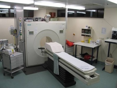
SPECT SCAN
A SPECT scan is a
type of nuclear imaging test, which means it uses a radioactive substance and a
special camera to create three-dimensional images that show how your organs
work.
 SPECT
is
an accurate aid in diagnosing Parkinson's Disease as it can show decreased
dopamine activity. Most SPECT scans involve two steps : receiving a radioactive dye and using
a SPECT machine to scan a specific area of the body. Before undergoing the SPECT
scan, patients receive a radioactive substance through an injection or through
an intravenous (IV) infusion into a vein in the arm. The health care team
position the patient on a table in the room where they undergo the SPECT scan. Most
scans can take 30 to 90 minutes. For more information go to
SPECT scan. SPECT
is
an accurate aid in diagnosing Parkinson's Disease as it can show decreased
dopamine activity. Most SPECT scans involve two steps : receiving a radioactive dye and using
a SPECT machine to scan a specific area of the body. Before undergoing the SPECT
scan, patients receive a radioactive substance through an injection or through
an intravenous (IV) infusion into a vein in the arm. The health care team
position the patient on a table in the room where they undergo the SPECT scan. Most
scans can take 30 to 90 minutes. For more information go to
SPECT scan.
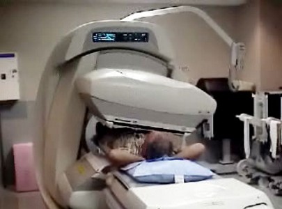
PET SCAN
The F-dopa PET scan is
an accurate aid in diagnosing Parkinson's Disease as it can show decreased
dopamine activity in the basal ganglia.
 Positron emission tomography (PET) is a nuclear medicine imaging technique
which produces a three-dimensional image or map
of functional processes in the body. It will take
approximately 30 to 60 minutes for radiotracer to travel through your body
and to be absorbed by the tissue being studied. You will then be moved into the PET
scanner and the imaging will begin. Actual scanning time is approximately 45 minutes. Images of tracer
concentration in 3-dimensional space within the body are then reconstructed by
computer analysis. For more information go to
PET scan. Positron emission tomography (PET) is a nuclear medicine imaging technique
which produces a three-dimensional image or map
of functional processes in the body. It will take
approximately 30 to 60 minutes for radiotracer to travel through your body
and to be absorbed by the tissue being studied. You will then be moved into the PET
scanner and the imaging will begin. Actual scanning time is approximately 45 minutes. Images of tracer
concentration in 3-dimensional space within the body are then reconstructed by
computer analysis. For more information go to
PET scan.
SMELL TESTS
The SIT, which is also known as UPSIT, consists of four
self-administered test booklets, each containing ten stimuli for
smell.
 Loss of olfactory function (sense of smell) is
common in Parkinson's Disease, and so is sometimes used as a means of diagnosis.
Respondents pick from one of four
multiple choices. For more information go to
SIT. 'Sniffin' Sticks' is a test of
nasal chemosensory performance based on pen-like odour dispensing
devices. It comprises three tests of olfactory function, for odour
threshold, odour discrimination and odour identification. The
specificity of the 16-item identification test from Sniffin' Sticks
(SS-16) when used in Parkinson's Disease was 89% to 90%, and there
was a sensitivity of 81% to 85%. Loss of olfactory function (sense of smell) is
common in Parkinson's Disease, and so is sometimes used as a means of diagnosis.
Respondents pick from one of four
multiple choices. For more information go to
SIT. 'Sniffin' Sticks' is a test of
nasal chemosensory performance based on pen-like odour dispensing
devices. It comprises three tests of olfactory function, for odour
threshold, odour discrimination and odour identification. The
specificity of the 16-item identification test from Sniffin' Sticks
(SS-16) when used in Parkinson's Disease was 89% to 90%, and there
was a sensitivity of 81% to 85%.
TRANSCRANIAL SONOGRAPHY
Transcranial Sonography is a non-invasive,
diagnostic technique that makes use of sound waves to create a digital image.
 Sound waves are produced by a transducer. Strong, short electrical
pulses from the ultrasound machine make the transducer ring at the desired
frequency. Materials on the face of the transducer enable the sound to be
transmitted into the body. The sound wave is partially reflected
from layers between different tissues. The return sound wave vibrates the
transducer, which turns the vibrations into electrical pulses. The electrical
pulses then travel to the
scanner where they are then transformed into a digital image.
For more information go to
Transcranial Sonography. Sound waves are produced by a transducer. Strong, short electrical
pulses from the ultrasound machine make the transducer ring at the desired
frequency. Materials on the face of the transducer enable the sound to be
transmitted into the body. The sound wave is partially reflected
from layers between different tissues. The return sound wave vibrates the
transducer, which turns the vibrations into electrical pulses. The electrical
pulses then travel to the
scanner where they are then transformed into a digital image.
For more information go to
Transcranial Sonography.
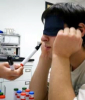
EYEBRAIN TRACKER
The eye-tracking
system, the Mobile Eye Brain Tracker (EBT), is available for the detection of
Parkinson-plus diseases.
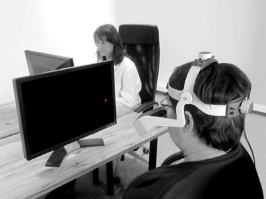 Parkinson-plus syndromes have additional features that
distinguish them from Parkinson's Disease.
Different areas of the brain are
involved in producing eye movements, and abnormalities that occur can be linked
to dysfunction in certain areas of the
brain. Results have shown that eye movements provide a more accurate early
diagnosis than traditional clinical examinations. The Mobile EBT is
non-invasive and costs less than regularly used imaging techniques.
For more information go to
Eye Brain Tracker. Parkinson-plus syndromes have additional features that
distinguish them from Parkinson's Disease.
Different areas of the brain are
involved in producing eye movements, and abnormalities that occur can be linked
to dysfunction in certain areas of the
brain. Results have shown that eye movements provide a more accurate early
diagnosis than traditional clinical examinations. The Mobile EBT is
non-invasive and costs less than regularly used imaging techniques.
For more information go to
Eye Brain Tracker.
PARKINSON'S KINETIGRAPH
The Parkinson�s KinetiGraph system consists of a sensor that is worn
around the wrist of the patient to record data about their symptoms.
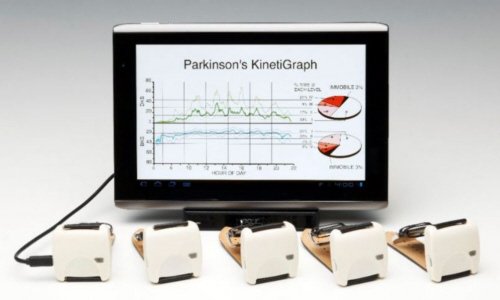 The
system also uses
a computer unit which receives that data and analyses it. The
device remotely records data about a person�s movement and via
algorithms, provides a report for the their neurologist showing an
objective measure of the presence and severity of bradykinesia and
dyskinesi. The device also reminds the person when to take their
Parkinson�s Disease drugs as prescribed by their medical
practitioner. For more information go to
Kinetigraph. The
system also uses
a computer unit which receives that data and analyses it. The
device remotely records data about a person�s movement and via
algorithms, provides a report for the their neurologist showing an
objective measure of the presence and severity of bradykinesia and
dyskinesi. The device also reminds the person when to take their
Parkinson�s Disease drugs as prescribed by their medical
practitioner. For more information go to
Kinetigraph.
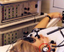
WEARABLE SENSOR
Mercury
is a wearable wireless sensor being developed as a means of enabling home
monitoring of the motion of people with Parkinson's Disease.
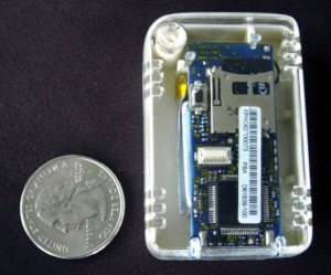 Patients wear
wireless nodes equipped with sensors for monitoring their movement and
physiological conditions. The basic approach is to capture data from each limb
using wearable sensors. The patient wears up to eight sensors and recharges the
sensors at night. A laptop in the patient's home stores the data. Data is then
sent via the Internet to the clinic where it is processed. Signals are subject to
extensive processing to evaluate the patient�s motor function.
For more information go to
Mercury Patients wear
wireless nodes equipped with sensors for monitoring their movement and
physiological conditions. The basic approach is to capture data from each limb
using wearable sensors. The patient wears up to eight sensors and recharges the
sensors at night. A laptop in the patient's home stores the data. Data is then
sent via the Internet to the clinic where it is processed. Signals are subject to
extensive processing to evaluate the patient�s motor function.
For more information go to
Mercury
 |
THE
COMPREHENSIVE GUIDE TO PARKINSON'S DISEASE
Keith Bridgeman, Tahira Arsham
The Comprehensive
Guide to Parkinson's Disease, which is fully referenced, and nearly
800 pages long, is the most comprehensive book concerning
Parkinson's Disease ever written. It includes its history, famous
people with Parkinson's Disease, its complete biochemisty, cytology
and cytological effects, anatomy and anatomical effects, physiology
and physiological effects, symptoms (of every system in the body),
diagnosis methods (observational, technological, chemical),
biochemical causes, toxic causes, genetic causes, pharmacological
causes, medical causes, treatments (biochemical, pharmacological,
surgical, natural, exercise methods, technological methods),
organisations, web sites, nursing.
CLICK HERE FOR MORE DETAILS |

|
.gif)
.gif)